Much of the total volume of your brain and spinal cord is made up of cells that support the nerve cells (neurons) in various ways but which don't carry information themselves. The collective name for these support cells is glial cells. Glia comes from the Greek word for glue and one of the roles of these cells is to hold the nerve cells in place.
Glial cells
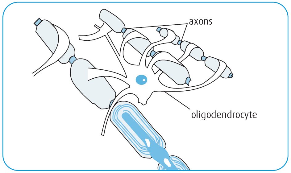
Oligodendrocytes
The glial cells that produce myelin in the central nervous system (your brain and spine) are called oligodendrocytes. In the peripheral nervous system, the myelin producing cells are called Schwann cells.
Each oligodendrocyte can supply myelin for the axons of several nerve cells and each axon can be supplied by several oligodendrocytes. The myelin produced by oligodendrocytes wraps around the axons in thin sheets like a Swiss roll. The myelin insulates the nerve axon so that electrical signals can be passed along the axon quickly and reliably.
Astrocytes
There are around five times as many astrocytes in your body as nerve cells. Under normal conditions, they line the blood-brain barrier (BBB) evenly, and help to keep it intact. Astrocytes also connect to nearby neurons and oligodendrocytes, regulating them and providing them with energy. Damage to a neuron makes nearby astrocytes reactive. They then tend to multiply and clump together, which may also make the BBB leaky.
Astrocytes secrete a variety of substances, some pro-inflammatory and some anti-inflammatory. In MS, reactive astrocytes both attract immune cells to a damaged nerve, and also surround and contain the active lesion. Astrocytes are involved in making the actual lesion scar, which prevents the damage spreading, but also prevents neural repair.
Microglia
Microglia are mobile cells that roam the CNS cleaning up damaged cells and pathogens. In MS and other conditions, they can be activated by inflammation, and become overactive. This may be how lesions get started.
Microglia may be particularly relevant to understanding the progressive forms of MS. In progressive MS, scans show active microglia at the edges of lesions. It is thought they may be nibbling away at the edges of lesions, enlarging them and causing disability accumulation.
Find out more
-
The role of astrocytes in multiple sclerosis
Frontiers in Immunology 2018 19(9) 217
Full article (link is external)
Microglial activation and its implications in the brain diseases.
Curr Med Chem. 2007 14(11):1189-97.
Summary (link is external)
Evaluation of Microglial Activation in Multiple Sclerosis Patients using Positron Emission Tomography
Frontiers in Neurology 2018 9:181
Full article (link is external)


