
Questions about MS?
MS Trust's Helpline offers free, confidential information and support for people affected by multiple sclerosis in the UK. Call 0800 032 38 39, email helpline@mstrust.org.uk or WhatsApp 01462 536008.
A lesion is an area of damage in the brain or spinal cord. Lesions in MS are sometimes also called plaques. They occur when the immune system attacks the myelin sheath around nerves. This causes inflammation and scarring, also known as sclerosis.
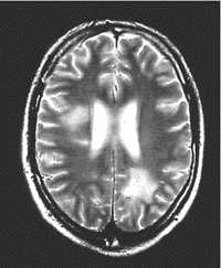
An MRI scan can detect MS lesions and also show the difference between active and non-active lesions. Active lesions show up in the scan as white patches when a contrast fluid containing gadolinium is injected. The gadolinium highlights the areas where inflammation is happening. If the lesion does not light up, then it is likely to be an older lesion, and more than 3 months old.
An MRI scan using gadolinium is also known as an MRI scan with contrast.
The presence, distribution and type of lesions seen in an MRI scan are useful clues for a neurologist to be able to give a diagnosis of MS. They form an important part of the McDonald Criteria that is used to make a diagnosis.
Not all lesions seen in an MRI are caused by MS. Lesions can be caused by other diseases such as migraine or stroke, and they can also develop with age. They can be detected in MRI scans before you experience any symptoms. Sometimes, a lesion might be detected in a scan that was done for another reason. You might then be given a diagnosis of RIS or radiologically isolated syndrome.
In some cases, neurologists can link a symptom that you have to a particular lesion in a scan, but this is not always possible. The size and position of a lesion on a scan does not always relate easily to your symptoms or experiences with MS. However, with regular scans, a neurologist may be able to tell how active your MS is, and how you may be affected.
Sometimes, lesions will repair themselves and will not be seen on subsequent scans. Persistent lesions may eventually show up as 'black holes', where the underlying neuron has suffered damage that cannot be repaired.
Some lesions get smaller or heal over time. Other lesions do not heal and tend to grow slowly over time. When looked at in MRI scans, the growing lesions have a dark rim of activity at the edges of the damaged area. They are known as chronic active lesions or smouldering lesions. In research they may be called paramagnetic rim lesions.
Smouldering lesions are thought to contribute to disease progression and worsening symptoms in MS. The dark rim seen in powerful MRI scans is caused by microglia, cells that normally roam the central nervous system cleaning up damaged cells. In MS, they may be activated by the immune system attack on nerve cells, becoming overactive and leading to further nerve cell damage.
Researchers found that people with four or more smouldering lesions were more likely to have developed progressive MS, and had cognitive and mobility problems from an earlier age, when compared to people with no smouldering lesions. They found that 56% of the people in their study had at least one smouldering lesion, regardless of the type of MS they had.
The concept of smouldering MS is guiding current research into medicines for MS. Current disease modifying drugs target the initial inflammatory attack on the nerves. Like firefighters at a forest fire, they put out the obvious blaze in the trees, but don't stop the smouldering burn happening under the leaves on the ground.
Newer drugs in development for MS aim to target the smouldering burn, stopping the microglia from enlarging existing lesions and letting them heal.

